Introduction
-
Retention of 18F FDG in the tumor reflects cellular growth and proliferation only in the past.
-
Moreover, 18F FDG uptake has also been associated with false positive findings resulting from unspecific tracer uptake in inflammatory process.
-
Many drugs have been designed to inhibit cell proliferation and/or to induce apoptosis.
-
Therefore in-vivo assessment of proliferation seems a promising tool for imaging cancer and assessing response to treatment.
-
Proloiferation imaging helps in
- Differentiation between benign and malignant.
- Non invasive grading.
18F FLT
- 3’deoxy-3'-18F Fluorothymidine (FLT) is used for noninvasive assessment of proliferation and more specific tumor imaging.
- It is a neucleoside analogue, derived from cytostatic drug Azidothymidine (AZT).
- There is upregulation of DNA synthesis pathway in tumors.
- Thymidine kinase 1 is idenitifed as key enzyme responsible for intracellular trapping 18F FLT. There is upregulation of TK-1 expression in tumors.
- It is taken up by the tumor cells by passive diffusion and facilitated transport (ENT-1)
- Imaging of DNA proliferation is superior to imaging glucose metabolism for assessing response to treatment because cells may maintain their oxidative and glycolytic function on spite of impaired DNA synthesis.
Applications:
Head and Neck:
-
FLT does not cross the intact blood-brain barrier, the physiological brain uptake is low, especially in comparison with 18F FDG.
-
Breakdown of the blood-brain barrier, however, can cause significant 18F FLT retention without cell proliferation.
-
Used in detection and treatment monitoring og gliomas.
-
It cannot differentiate between high grade and low grade tumors.
-
Physiological uptake in neck region is low. Therefore 18F FLT is believed to discriminate between inflammatory and neoplastic lymph node involvement. But false positives is seen in some lymph nodes.
-
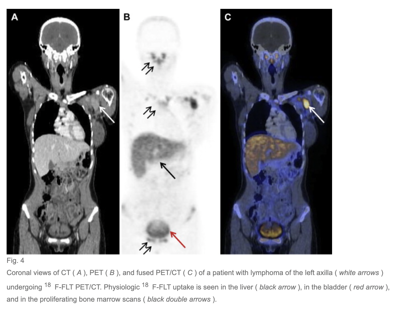
Further physiologic uptake is noted in proliferating bone marrow present in the cervical spine and to a lesser extent the skull. Therefore, there is decreased sensitivity in bone marrow infiltration and primary and secondary bone lesions.
-
However, in patients with NHL, even osseous lesions were detected because focal FLT uptake was greater than in surrounding bone marrow.
-
High physiologic uptake in proliferating bone marrow is successfully used to visualise the viable bone marrow compartment in patients with aplastic anemia and to identify organs with ongoing extraosseous hematopoiesis.
Thorax:
- Physiologic lung parenchyma shows only ver low 18F FLT uptake, and therefore permits the proposal that any kind of increased pulmonary 18F FLT uptake is highly suspicious of a malignant tumor.
- Increased FLT uptake in heart is because of accumulation of the tracer in the blood pool and muscle tissue of LV, obscuring breast and lung lesion.
- However, blood pool activity is washed out by 45 minutes.
- Physiologic uptake is also noted in the rib cae and dorsal vertebrae due to proliferating bone marrow.
Abdomen:
-
Increased physiologic uptake is noted in liver, intestines, ribs, dorsal and lumbar vertebrae.
-
18F FLT undergoes extensive glucoronidation in the human liver, resulting in approximately 25% of plasma activity being present as 18F FLT glucoronide at 60 minutes after injection.
-
Even if high uptake is noted in the liver, suspected HCC lesion shows higher uptake than surrounding liver activity.
-
Attempts to reduce the liver uptake using probenecid were done, but failed.
-
Physiologic uptake in GI mucosa is due to mucosa cells undergoing rapid proliferation.
Pelvis:
- FLT is excreted by kidneys in its non metabolised form and also in 18F FLT glucoronide form.
- At 60 minutes post injection, 18F FLT is seen in kidneys and genitourinary system.
- No physiologic uptake is noted in uterus, ovaries, prostate and seminal vesicles.
Treatment induced effects:
- In response to chemotherapy, reduced FLT uptake is expected.
- But sometimes, increased FLT uptake is noted, due to flare phenomenon.
Follow me on Twitter @KoteRutuja for more updates and resources.
References
Herrmann K, Buck AK. Proliferation imaging with ¹⁸F-fluorothymidine PET/computed tomography: physiologic uptake, variants, and pitfalls. PET Clin. 2014 Jul;9(3):331-8. doi: 10.1016/j.cpet.2014.03.005. Epub 2014 Apr 26. PMID: 25030396.