- Paget disease is often included in the metabolic bone disease category, although mechanisms causing this disorder are not entirely understood, with genetic and environmental causes proposed.
- A chronic disease of the elderly.
Clinical Features:
Patients may experience
- pain,
- arthritis, and
- neurological symptoms related to changes in the bone.
- Congestive heart failure can occur
- rarely (1%) patients develop osteosarcomas.
Phases:
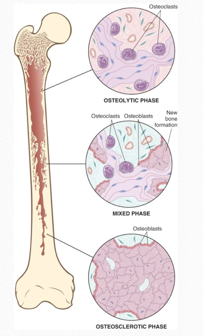
The disease has three phases:
- the early resorptive,
- mixed middle, and
- final sclerotic phases.
Investigations:
- The diagnosis usually can be made by radiographs, which reveal lytic lesions in early cases and coarsened, expanded bones as the disease progresses to the final phase.
- Although CT and MR can be used to assess complications, bone scan is highly sensitive and useful to evaluate the extent of disease.
Bone scan:
- Increased uptake is seen in all stages of untreated disease, although sensitivity is lower in the early lytic stage.
- Activity decreases with effective therapy.
- In addition, the patterns of Paget disease must be recognized because it may be found incidentally because many patients are asymptomatic and undiagnosed.
- Bone often appears expanded.
- When the tibia is involved, bowing is often seen, and in the spine, fractures can happen.
- The pelvis is the most commonly involved site, followed by the spine, skull, femur, scapula, tibia, and humerus.
Osteoporosis circumscripta
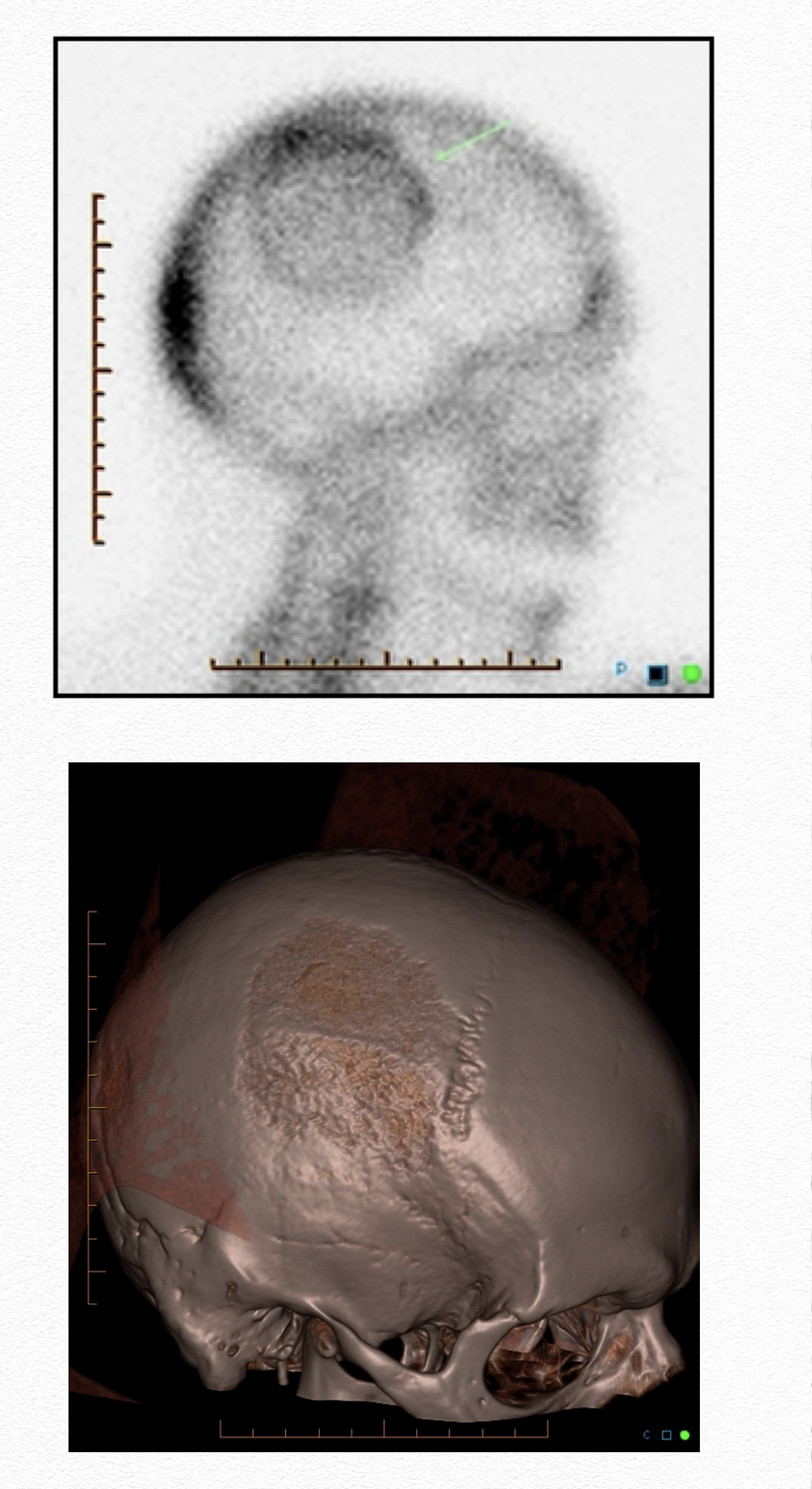
- A characteristic rim of increased uptake borders the lesion.
- Lytic phase/lesion involving skull bones
Mickey Mouse sign
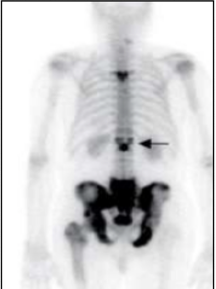
- Characteristic appearance of vertebral Paget’s disease
- Abnormal tracer accumulation throughout the vertebra
- Affecting body and posterior elements
- Clover/heart/Mickey Mouse sign
Lincoln sign
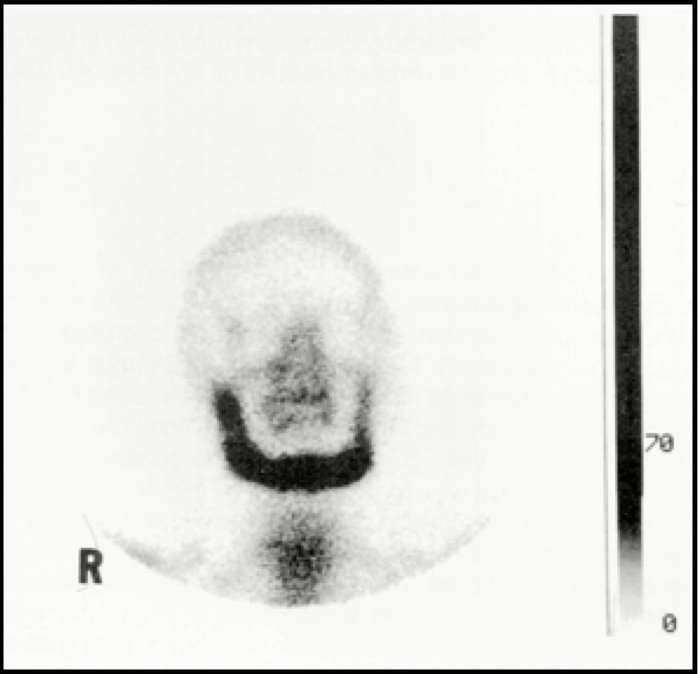
- monostotic Paget’s of mandible
Short pant sign
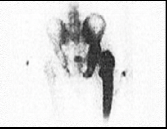
- Lower spinal, pelvic and proximal femoral involvement