- The parathyroid glands are able to concentrate a variety of chemical substances.
- Tc99m sestamibi is the most commonly used agent.
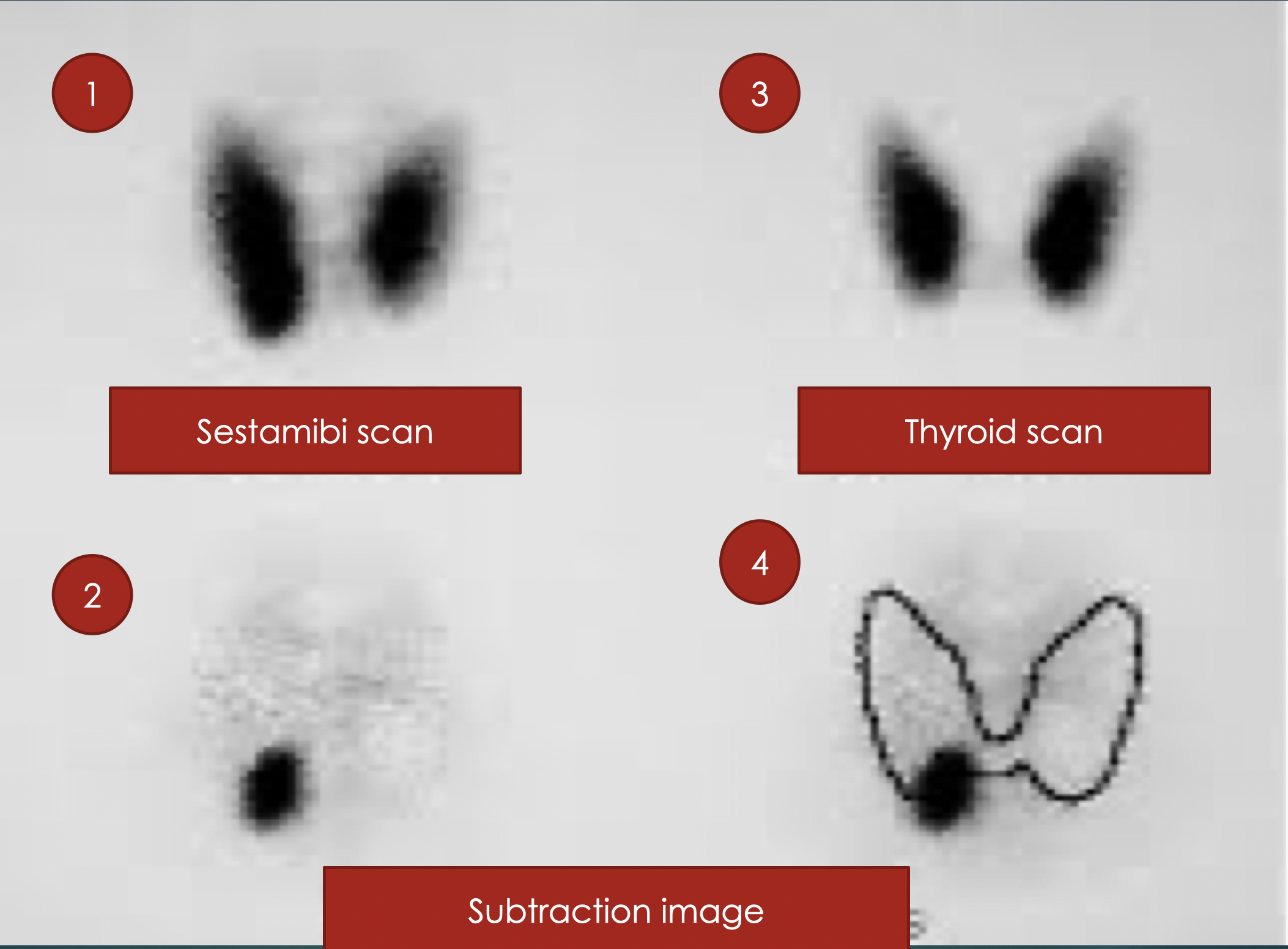
- Works on differential washout of MIBI from thyroid and parathyroid gland.
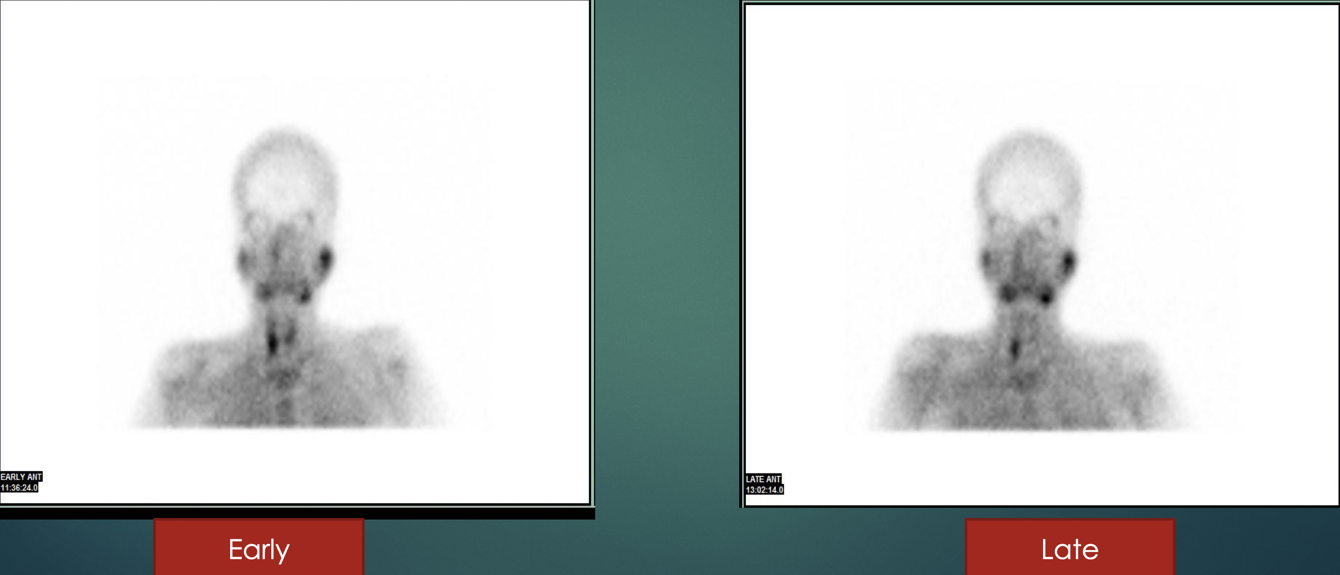
- Single isotope- double phase technique
- MIBI washes out early from the thyroid gland compared to parathyroid gland.
- Retention is proportional to number of oxyphil cells.
- Physiological uptake noted in thyroid gland, heart, liver, salivary gland.
Protocol
- Single injection of 20-30mCi of Tc99m Sestamibi given I.V.
- Planer image is taken at 15 minutes (early) and 1.5 to 3 hours later (late)
- Followed by SPECT or SPECT/CT for each time.
- Thyroid scan with Tc99m pertechnetate is done
- Chest/whole body survey is done to look for ectopic lesion/ brown tumour.
- Subtraction images are taken and compared
- A persistent focus of activity, in delayed images, relative to the thyroid gland activity, indicative of the parathyroid lesion.
Advantages:
- Relatively operator independent in comparison with USG
- Detects ectopic and posterior glands that USG may miss
- Provide valuable anatomic landmark
- Lower radiation dose than with 4D CT
- Can we used as a tool in operating room for parathyroid localization- Radio guided parathyroidectomy.
Limitations:
- Patients must remain relatively motionless for a longer period of time
- More expensive than USG and 4D-CT scans
- Scintigraphy perform poorly at detecting small glands (<300mg), multi-gland disease (<50% sensitivity)
- Subtypes with fewer oxyphil cells do not take up tracer
- Sometimes parathyroid carcinoma, thyroid nodules, thyroid malignancy lymph adenopathy can also mimic these findings.