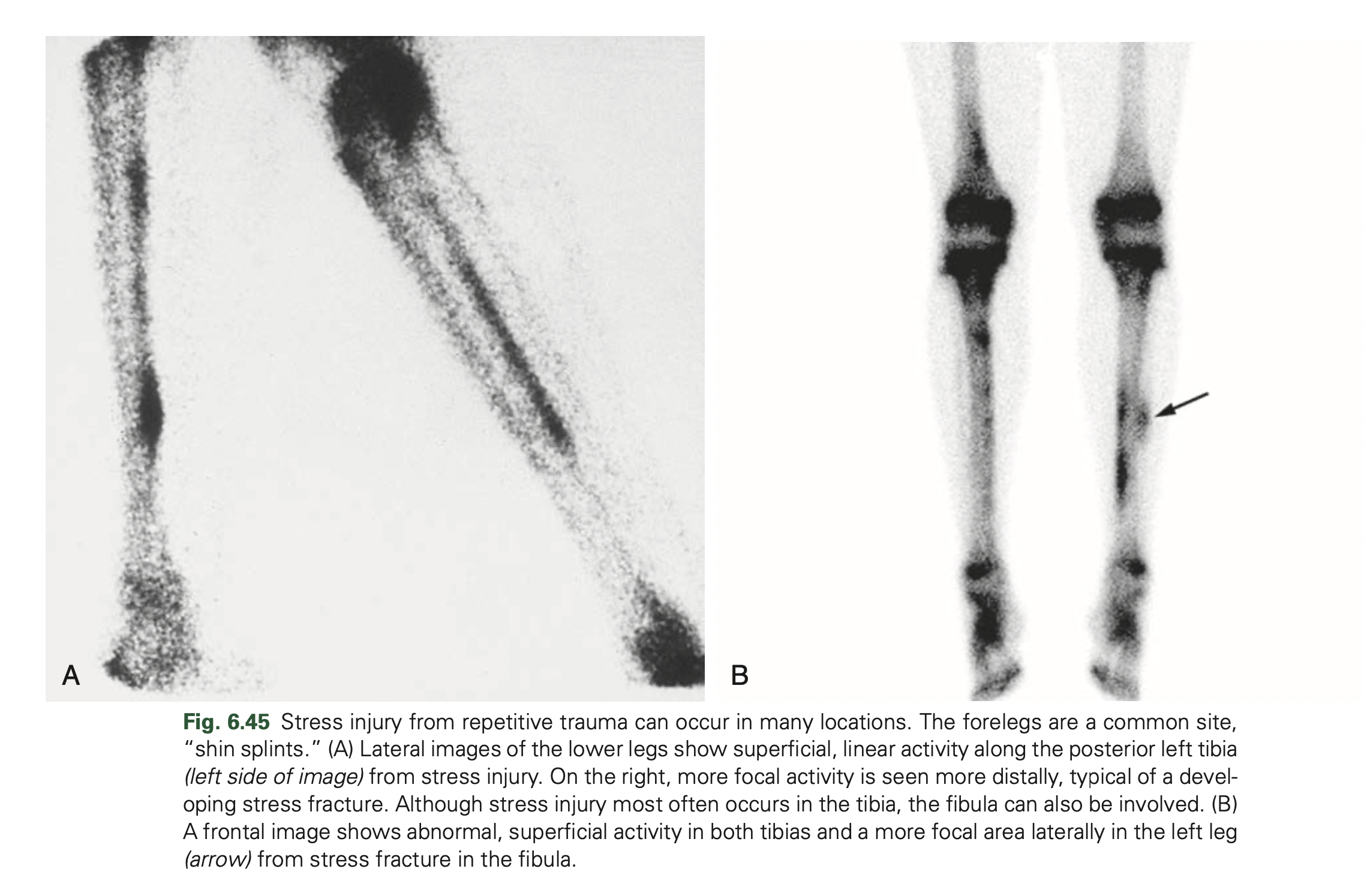
A significant change in activity level or a repetitive activity may lead to injury to the bone. This may be seen in “shin splints,” a term applied generically to describe stress-related leg soreness along the medial or posteromedial aspect of the tibia.
DEFINITION:
In nuclear medicine, the term is used for a specific combination of clinical and scintigraphic findings: peripheral, linear tracer uptake is seen on the scintigram, typically involving a large portion of the middle to distal tibia. If the process causing injury is allowed to continue to the point of overt fracture, healing predictably takes several months or more, compared with the several weeks required for healing of an early stress reaction.
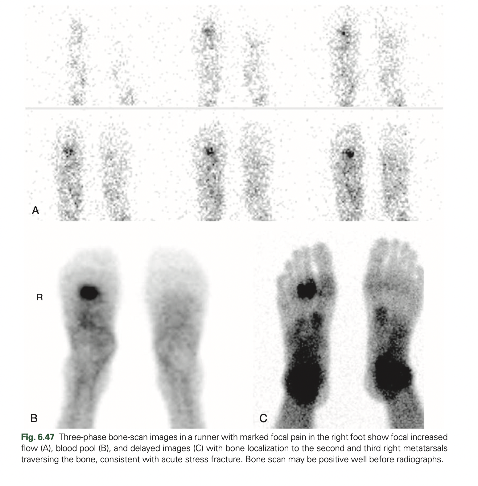
Therefore prompt diagnosis and appropriate change in activity are critical. Bone scan is exquisitely sensitive to detect Stress fracture.
IMAGE FINDINGS
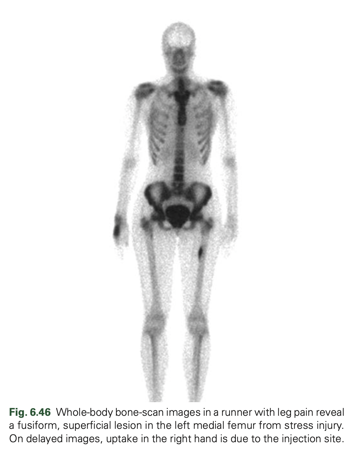
- Skeletal scintigraphy reveals characteristic intense uptake at the fracture site ranging from the earlier oval or fusiform uptake to activity traversing the bone in outright fracture Stress fracture can become positive on all three phases of the bone scan and findings are often seen before radiographs reveal the cortical thickening in injury and fracture lines as disease progresses.
- Stress injury is not uncommonly multifocal; thus additional sites of involvement may be detected.
- A phenomenon perhaps related to shin splints is activity induced enthesopathy.
- In athletes, repeated microtears with subsequent healing reaction can result in increased tracer uptake at the site of tendon or ligament attachment.
- Osteitis pubis, plantar fasciitis, Achilles tendonitis, and some cases of pulled hamstring muscles are examples.
- A periosteal reaction develops at the site of stress, sometimes resulting in increased skeletal tracer localization.
- Stress injuries can also occur in the spine.
- Spondylolysis occurs in the lumbar spine in the pars articularis, often seen as a result of repetitive trauma in young athletes.
- Most commonly the abnormality occurs at L4 to L5.
- In some instances, all examinations, including radiographs, MR, and planar bone scan, may be normal, but a SPECT study may reveal the pathological condition.
- When available, SPECT/CT can provide an optimal assessment.
Follow me on Twitter @KoteRutuja for more updates and resources.